PEOPLE
BPI Team
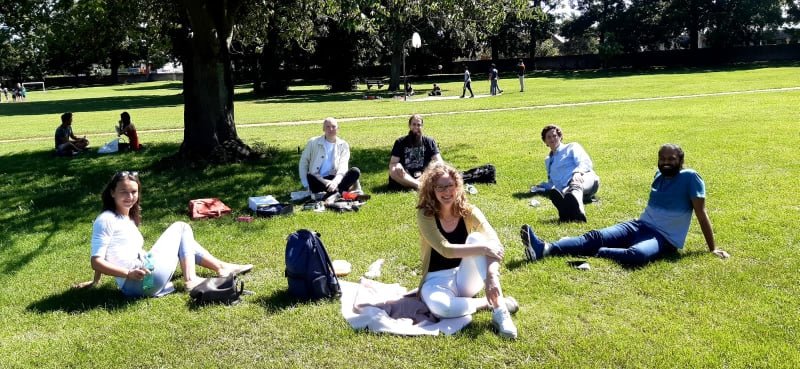
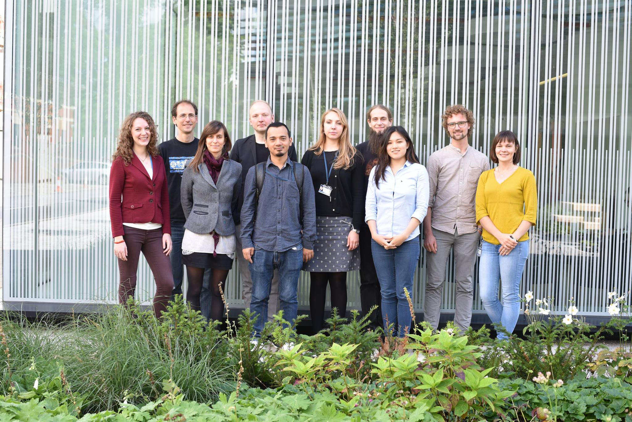
Huw Colin-York, Veronika Pfannenstill, Anna Schepers, Jacky Ka Ko Long, Narain Karedla, Kaitlyn Purdie, Jess Stone, Shuyang Xu, Marco Fritzsche.
Marco Fritzsche
Principal Investigator
Email: marco.fritzsche@kennedy.ox.ac.uk
Phone: +44 (0) 1865 612685
Biomedical sciences increasingly recognise the physics of immunity in health and disease. For the most part, this is due to an emerging new perspective of the broad impact of mechanobiology on the immune response. The BPI Lab aims to understand the biological significance of mechanobiology for immunophysiology. For this mission, we develop advanced technology, microscopy, and analysis approaches.
BSc (2003-2006) in Mathematics, Department of Applied Mathematics, University of Saarbrücken, Germany.
BSc (2003-2006) in Physics, Department of Theoretical Physics, University of Saarbrücken, Germany.
MSc (2006-2008), Karsten Kruse, Theoretical Physics, University of Saarbrücken, Germany.
PhD (2008-2012), Guillaume Charras, Experimental Physics and Biology, University College London, UK.
Postdoc (2013-present), Christian Eggeling, Weatherall Institute of Molecular Medicine, University of Oxford, UK. Visiting Researcher (2015-2016), Eric Betzig, Janelia Farm, Howard Hugh Medical Institute, USA.
Principal Investigator (2016-2020), MRC Junior Principal Investigator, MRC Human Immunology Unit, Weatherall Institute of Molecular Medicine, and Kennedy Institute for Rheumatology, University of Oxford, UK. Principal Investigator (2020-Present), Associate Professor, Rosalind Franklin Kennedy Trust Research Fellow, Kennedy Institute for Rheumatology, University of Oxford, and Rosalind Franklin Institute, UK. Scientific Director (2021-Present), Oxford-ZEISS Centre of Excellence between Institute of Developmental and Regenerative Medicine and Kennedy Institute for Rheumatology, University of Oxford, UK. Full Professor (2023-Present) of Biophysical Immunology, Kennedy Institute for Rheumatology, University of Oxford, UK.
Kseniya Korobchevskaya, PhD
Advanced Microscopy Manager
Email: kseniya.korobchevskaya@kennedy.ox.ac.uk
Having a strong background in experimental physics and optics, I was always interested in biological applications. For that reason, after completing my PhD in ultrafast spectroscopy I moved to the field of super-resolution microscopy. As a part of the Nanoscopy group at IIT I worked on the development of label-free near IR pump-probe nanoscope. Besides, I was also closely involved in other projects related to nonlinear microscopy, such as subtraction microscopy approaches for second-harmonic generation imaging of collagen fibres or two-photon spectrofluorimetric characterization of a new set of red-emitting proteins. I joined the BPI Lab to develop the eTIRF-SIM.
Presently, I am working in the BPI Lab and am an Advanced Microscope Manager in the Oxford-ZEISS Centre of Excellence.
PhD (2010-2013), Alberto Comin, Ultrafast Spectroscopy, Italian Institute of Technology, Genova, Italy.
Postdoc (2013-2016), Alberto Diaspro, Nanoscopy group, Italian Institute of Technology, Genova, Italy.
Microscopy Staff Scientist (2016-2021), Marco Fritzsche, Kennedy Institute for Rheumatology, University of Oxford, UK. Advanced Microscopy Manager (2021-Present) Oxford-ZEISS Centre of Excellence between Institute of Developmental and Regenerative Medicine and Kennedy Institute for Rheumatology, University of Oxford, UK.
Helena Coker, PhD
Microscopy Staff Scientist
Email: helena.coker@kennedy.ox.ac.uk
Microscopy is the key to capturing the biological events which are important to researchers and to the community. Microscopists are necessarily moving away from the turn-key workhorses, towards more specialised and individualised instruments, as the limits and rules that governed light microscopy are continually pushed. I find a lot of joy in figuring out these new or little used technologies, then condensing the need-to-knows and how-tos such that they are accessible, and researchers can make the most of them.
MSc (2009-2014) Natural Sciences, University of Bath, UK. DPhil. (2014-2018) ‘Anomalous Diffusion in Artificial Lipid Bilayers’ Physical and Theoretical Chemistry, University of Oxford, UK. Imaging Specialist (2018-2021) Lattice Light Sheet Microscopy, University of Warwick, UK. Advanced Microscopy Manager (2021-Present) Oxford-ZEISS Centre of Excellence between Institute of Developmental and Regenerative Medicine and Kennedy Institute for Rheumatology, University of Oxford, UK.
Huw Colin-York, PhD
Postdoctoral Researcher
Email: huw.colinyork@kennedy.ox.ac.uk
The role of mechanical force has gained increasing interest in the field of cell biology. This has come from the realisation that cells are continually subject to stresses and strains induced by the cellular environment. Cells are known to be able to sense and react to forces imposed on them by their local environment as well as being able to directly impart force during motility and adhesion. This is also true for cells of the immune system, specifically, T-cells are known to be highly motile, navigating through the blood, tissue and lymphatic system. To achieve this level of mobility, T-cells rely on a highly dynamic cytoskeleton. On encountering an Antigen Presenting Cell (APC) that is presenting a foreign antigen, T-cells become activated. This highly selective process by which a T-cell is able to bind, recognise and react to only foreign antigens has been the focus of intense study due to its crucial importance in the adaptive immune response. Despite this, the early stages of T-cell activation remain unclear. The possible active role mechanical force might play in this process has been largely overlooked and a quantitative approach to measuring the forces generated during this interaction is lacking. By employing tools from biophysics and cell mechanics the work proposed aims to investigate the possible role of mechanical force in the initiation and continuation of the T-cell-APC interaction.
Narain Karedla, PhD
Postdoctoral Researcher
Email: narain.karedla@chem.ox.ac.uk
Optical microscopy and spectroscopy have witnessed an enormous growth in recent years and have become indispensable tools in physical and live science applications. Novel techniques that allow quantitative deep tissue imaging with sub-cellular resolution and super-resolution techniques that can achieve nanometer resolution are powerful means for studying biological processes. I am an experimental physicist by training with a deep motivation in developing and exploring new advanced optical tools. During my PhD, I have developed and worked on several single molecule spectroscopy and super-resolution techniques for biophysical applications. One notable example is metal-induced energy transfer (MIET) that is capable of localizing individual fluorescent emitters with isotropic 3D nanometer resolution. During my postdoctoral training, I built a new method for quantifying electrostatic surface charge density of solids immersed in fluids using optical microscopy. This method also allows for the direct measurement of effective charges of biomolecules in solution, a fundamental characteristic that is crucial to study ligand-receptor binding interactions and structure-charge interrelationship of biomolecules.
Driven by the motivation to design and deliver advanced optical microscopy methods, I am currently building a lattice light sheet microscope coupled with structured illumination and adaptive optics in the BPI lab. This transformative technology will allow acquisition of 3D super-resolution volumetric data at one to two orders of magnitude faster than a conventional widefield or confocal microscope. The microscope will be a powerful tool to interrogate and correlate biophysical events within single cells and cells in their tissue environments. In particular, one of my main interests it to use the technology to unravel the dynamic biophysical processes spanning over multiple length- and time-scales that are involved during T-cell activation when interacting with their local microenvironment
Integrated MSc (2007-2012) Chemistry, Indian Institute of Technology, Roorkee, India.
Research Internship (2009), Naresh Patwari, Indian Institute of Technology, Bombay, India. Research Internship (2010), Dirk-Peter Herten, Ruprecht - Karls University, Heidelberg, Germany. Research Internship (2011), Thorsten Wohland, National University of Singapore, Singapore. Postdoc (2016- Feb 2018), Dr. Jörg Enderlein, Georg-August-Universität, Göttingen, Germany. Postdoc (Mar 2018 - Sept 2018), Shimon Weiss, Bar-Ilan University, Ramat Gan, Israel. Postdoc (Apr 2019 - Nov 2020), Madhavi Krishnan, Alexander von Humboldt Fellow, University of Oxford, Oxford, UK. Advanced Microscopy Specialist (2021 - Present), Marco Fritzsche, Rosalind Franklin Institute and Kennedy Institute for Rheumatology, University of Oxford, UK.
Jacky Ka Long Ko, PhD
Bioimage Analyst
Email: ka.ko@kennedy.ox.ac.uk
I am expertised in biomedical image processing with years of experience in developing and designing associate algorithms. I am responsible for deploying machine learning techniques in microscopic images and managing image analysis infrastructure at the Oxford-ZCoE. I also assist other team members to develop automated analytical pipelines for the interpretation of microscopy data, including feature detection, segmentation, and motion tracking
BSc (2010 - 2013) in Physics, The Chiniese University of Hong Kong, Hong Kong
MPhil (2015 - 2017) in Imaging and Interventional Radiology, The Chiniese University of Hong Kong, Hong Kong
PhD (Sept 2019 – Aug 2022) in Imaging and Interventional Radiology, The Chiniese University of Hong Kong, Hong Kong Bioimage Analyst (Nov 2022– Present), Marco Fritzsche, Oxford-ZEISS Centre of Excellence between Institute of Developmental and Regenerative Medicine and Kennedy Institute for Rheumatology, University of Oxford, UK.
Anna Schepers, PhD
Post Doctoral Researcher
Email: anna.schepers@kennedy.ox.ac.uk
Immune cells must orchestrate a complex series of morphological changes, along with biochemical and biomechanical signaling events, while constantly being exposed to the mechanical constraints of their environment. I am interested in understanding how the mechanical properties of cellular components, individual cells, and their surrounding environment are linked to cellular function. To explore this, I use various techniques to quantify protein and cell mechanics. During my fellowship in the BPI lab, I am focusing on how the stiffness of target cells influences the T cell response, and how this response is affected by confinement.
BSc (2011 - 2014) Nanoscience, University of Hamburg, Germany.
MSc (2014 - 2017) Nanoscience, University of Hamburg, Germany 2017: Jean-Marie Lehn and Klaus von Klitzing Prize for the best M.Sc. thesis in Nanoscience.
PhD (2017 – 2021) Physics of Biological and Complex Systems, University of Goettingen, Germany, 2022: Jan Peter Toennies physics prize for outstanding PhD theses in Experimental Physics.
Postdoc (2022– 2024), Marco Fritzsche, Rosalind Franklin Institute and Kennedy Institute for Rheumatology, University of Oxford, UK.
Postdoc (2024-Present), Marco Fritzsche, Walter-Benjamin Scholarship, Kennedy Institute for Rheumatology, University of Oxford, UK.
Veronika Pfannenstill, DPhil
Post Doctoral Researcher
Email: veronika.pfannenstill@stx.ox.ac.uk
Throughout my studies, I have developed a profound interest in problems at the interface between physics, biology, and medicine. This is due, in part, to my fascination in seeing how fundamental research and abstract concepts originating from physics, hand in hand with novel experimental tools, explain or at least help to improve our understanding of living systems. Witnessing the tremendous technological and conceptual progress made over the last decade, I am convinced that now is a right moment to address some of the essential questions in the field of biophysics. I have thus decided to contribute to this endeavor and continue my scientific development accordingly.
Currently, I am working on the development of a novel high-throughput fluorescence microscopy platform that can detect and track the ability of immune cells to kill pathogens within large cell ensembles at single-cell sensitivity. This technology has the potential to advance our understanding of the key mechanisms underlying the human immune response.
BSc (2013 - 2016) in Technical Physics, Technische Universität Wien, Austria. BSc internship (summer 2015) Friedrich-Alexander Universität Erlangen-Nürnberg, Germany. MSc (2016 - 2019) in Technical Physics, Technische Universität Wien, Austria.
MSc internship (Feb - Jul 2018) Laboratory of Physics of Living Matter, École Polytechnique Fédérale de Lausanne (EPFL), Switzerland.
DPhil (Oct 2019 – Jul 2024), Marco Fritzsche, Kennedy Institute for Rheumatology, University of Oxford, UK.
Postdoc (Aug 2024 – Present), Marco Fritzsche, Kennedy Institute for Rheumatology, University of Oxford, UK.
Kaitlyn Purdie, MSc
DPhil student
Email: kaitlyn.purdie@lmh.ox.ac.uk
I completed my undergraduate degree in Biology at the University of York where I gained experience in mathematical modelling through my research project exploring the population dynamics of Leishmania spp. I am now completing my MSc in Integrated Immunology at the University of Oxford. Throughout both degrees I have become increasingly aware of the mechanistic principles that form the foundations of many aspects of biology, particularly in immunology. My interest in this interplay between biophysics and immunology led me to my current research project within the BPI lab where I am investigating the role of mechanobiological and signalling cues in the activation of primary human T cells. Beginning in September 2021, I will remain in Oxford to complete my DPhil in Molecular and Cellular Medicine which will be a collaboration between the BPI lab and the Nanchahal group. Here, the focus will be on developing novel microscopic methodologies to study the biomechanical behaviour of human liver stromal cells to quantify liver fibrosis and regeneration.
BSc (2015 - 2018) Biology, University of York, UK. MSc (2020 - 2021) Integrated Immunology, University of Oxford, UK. DPhil (2021 - Present) Molecular and Cellular Medicine, Marco Fritzsche, Kennedy Institute for Rheumatology, University of Oxford, UK.
Alumni
Yuexuan Zhang, MSc
Email: yz756@cam.ac.uk
During my studies, I developed a strong passion for utilizing various microscopy techniques in conjunction with classical biochemical methods to explore the dynamics and structures of key proteins involved in essential cellular processes. My undergraduate project on cancer cell migration sparked my enthusiasm for understanding the interplay between mechanical and chemical signalling in tumour development and progression.
During my summer project in the BPI lab, I focused on using autofluorescence lifetime imaging to investigate the complexity and heterogeneity of the colorectal tumour microenvironment. Additionally, I examined the lifetime of colorectal cancer cell lines in response to different drugs, aiming to establish cellular-level lifetime benchmarks. In my free time, I enjoy listening to Kseniya's jokes and discussing cell biology.
BSc, MSc (2021 - 2025) Natural Sciences (Biochemistry), University of Cambridge, UK
Ben Xu, MSc
Email: yiming.xu@exeter.ox.ac.uk
I am a MSc student in the Oxford Integrated Immunology program. I completed my BSc in Laboratory Medicine and Pathobiology at the University of Toronto in Canada. My MSc project in the BPI focused on the mechanobiology of chimeric antigen receptor (CAR) -T cell immune synapses. CAR immune synapses are structurally and functionally different from that of natural T cells, but their mechanobiology is less well studied. We used microsphere-based 3D traction force microscopy (TFM) to study the immune synapse of CD19 CAR-T cells, a key therapy for B cell malignancies. Coming from a biology background, my MSc project was a great opportunity to appreciate biophysical principles in immunology and advanced imaging techniques. After my MSc, I will take up a PhD position at the University of Geneva studying the role of monocytes in atherosclerotic plaques.
Mike Barnkob, MD, PhD
Postdoctoral Researcher
Email: mike.barnkob@kennedy.ox.ac.uk
I am a doctor specialising as a clinical immunologist, with a Ph.D. (Oxon 2018) in tumour-immunology. My research interests are chimeric antigen receptor (CAR) T cells, synthetic biology and cancer.
My research is focused on exploring the interactions between CAR T cells and tumours at both the immediate stage and long-term, and to use this knowledge to identify and develop novel CAR therapies against hematological cancers.
It’s my belief that the future of oncology lies at the interphase between synthethic biology and immunology. By reprogramming T cells to better function in tumours or to attack novel targets, we are able to increase the strength and power of the body’s own immune system, and hopefully cure cancer.
Marcel Issler, MSc
MSc Erasmus+ Trainee
Email: marcel.issler@kennedy.ox.ac.uk
My current research conjoins nouvelle microscopy and computational methods to elucidate the impact of integrins on the mechanical interaction during immunological synapse formation. Utilising astigmatic Traction Force Microscopy (aTFM) I aim to quantify the three-dimensional forces during this process with high spatio-temporal precision.
Currently I engineer ultra-soft hydrogels and quantify their performance with PIUMA/AFM to rigidify our experiments. Additionally, I create simulations for various traction force microscopy experiments to quantify the precision of our experiments.
BSc (2017-2021) in Biophysics, Humboldt University to Berlin, Germany BSc Internship (Autumn 2020) Swiss European Mobility Program (SEMP) Trainee at the Laboratory of Applied Mechanobiology, Eidgenössische Technische Hochschule Zürich (ETH Zurich), Switzerland MSc (2020-2023) in Biophysics, Humboldt University to Berlin, Germany MSc Internship (2022-2023) Erasmus+, Marco Fritzsche, Trainee in Biophysical Immunology, University of Oxford, UK
Falk Schneider, PhD
Postdoctoral Researcher
Email: falkschn@usc.edu; Twitter: @faldalf
Life is dynamic. Living cells rely on the versatile, coordinated and chaotic motions and interactions of a myriad of molecules. Studying this molecular motions and dynamic reorganisations requires high spatial and temporal resolution and cannot be achieved with conventional imaging approaches. I am a biochemist by training and naturally curious about molecular organisation and its impact on the larger length-scales. During my time in the BPI lab, I worked on extending the available fluorescence fluctuation based toolkit to study nano-scale plasma membrane organisation during T-cell activation. T-cell signalling starts at the membrane as this structure displays the first point of contact with the environment or target cell. Characterising its (pre-)organisation will provide means to understand initiation of signalling and how to tune it.
BSc (2010-2013) in Biochemistry, University of Hanover MSc (2013-2015) in Biochemistry, Hanover Medical School PhD (2015-2019) in Medical Sciences, University of Oxford PostDoc (2020-2021) with Prof. Marco Fritzsche (BPI lab) at MRC Human Immunology Unit, Weatherall Institute of Molecular Medicine, and Kennedy Institute for Rheumatology, University of Oxford, UK. PostDoc (2021 - present) with Prof. Scott Fraser (Fraser Lab - Translational Imaging Center) at University of Southern California, Los Angeles, United States
Liliana Barbieri, PhD
Postdoctoral researcher
Email: liliana.barbieri@dtc.ox.ac.uk
I am a physicist by training with strong interest in understanding the physical principles of cell-biological systems. Currently, I am working on the characterisation of the reorganisation dynamics of the cortical actin cytoskeleton during T-cell activation. Cortical actin dynamics give rise to active force production, which I quantify by simultaneous read-out of the mechanical forces and the underlying turnover dynamics of actin and key proteins involved in the activation process such as the T-cell receptor. Understanding the role of the actin cytoskeleton is now becoming a contentious questions in immunology but progress has previously been limited, mainly due to the use of conventional-resolution microscopy, which inevitably misses essential details due to limited resolution. To overcome these limitations, I will work with more advanced microscopy techniques, such as super-resolution optical nanoscopy combined with traction force microscopy and atomic force microscopy.
BSc (2009 - 2013) Physics, University of Palermo, Italy
MSc (2013 - 2016) Physics of matter, University of Palermo, Italy
Erasmus program (Sept 2015 - Jan 2016) in Exploration du Vivant et de l'Environnement, Université Joseph Fourier, Grenoble – France
MSc internship (Jan - Jun 2016) Institut de Biologie Structurale, Grenoble, France
PhD (Oct 2016 – May 2021) Oxford-Nottingham Biomedical Imaging, Marco Fritzsche, MRC Human Immunology Unit, Weatherall Institute of Molecular Medicine, and Kennedy Institute for Rheumatology, University of Oxford, UK. Postdoc (Apr 2021 - 2022), Marco Fritzsche, MRC Human Immunology Unit, Weatherall Institute of Molecular Medicine, and Kennedy Institute for Rheumatology, University of Oxford, UK. Bioimage Analyst (2022-Present), Oxford Nano Imaging, Oxford, UK.
Aurélien Barbotin, PhD
Staff scientist
Email: aurelien.barbotin@inrae.fr
After my PhD work in adaptive optics for STED microscopy, I joined the BPI lab to apply my skills in optics and image processing to solve biophysical problems in the field of immunology. I am particularly interested in understanding the spatiotemporal interactions underlying immunological processes. For this, I use in collaboration with the rest of the lab a combination of bespoke optical microscopes and customised image analysis algorithm to extract spatiotemporal information about calcium signalling and cell death.
BSc (2010-2012) Mathematics, Physics and Chemistry - Lycée Hoche, Versailles, France.
MSc (2012-2016) Optics - École polytechnique, France/ Institut d'Optique Graduate School, France.
PhD (2016-2020) Oxford-Nottingham Biomedical Imaging doctoral training programme, at the department of Engineering Science, University of Oxford, UK. Staff scientist (2020-2021) Bioimage analyst, Marco Fritzsche, University of Oxford, UK. Postdoc (2021-present) Marie Skłodowska-Curie fellow in the ProCeD team at Micalis Institute, Paris-Saclay, France.
Karim Ajmail, BSc
MSc student
Email: karim.ajmail@mtl.maxplanckschools.de
During my Erasmus+ research internship at the Kennedy Institute of Rheumatology, I took part in the development of a new microsphere traction force microscopy platform. Here we use the lattice lightsheet microscopy to record 3D traction forces, that T-cells exert to hydrogel microspheres. Currently, I am pursuing my masters at the Max Planck School in Göttingen.
Gabrielle Chappell, BSc
MSc student
Email: gabrielle.chappell@kellogg.ox.ac.uk
I am a current MSc Integrated Immunology student at the University of Oxford. In my undergraduate degree, I studied Mathematics and Biology at the University of St Andrews. My research background is in immuno-oncology, particularly in adoptive T-cell therapies including tumor-infiltrating lymphocytes (TIL) and chimeric antigen receptor T (CAR-T) cells. How T-cells discriminate between self and non-self, infected-self, or cancerous-self is poorly understood, yet it is a fundamental aspect of the adaptive immune response. Therefore, understanding how TCR-peptide affinity interactions and calcium signaling impact T-cell activation and immunological synapse formation has a breadth of translational applications. I will be pursuing a bioinformatics project with the BPI lab to help investigate and analyze how the quantitative strength of calcium signaling influences actin cytoskeletal rearrangement and immunological synapse formation. After completing my Masters, I will be undertaking a PhD at the Kennedy Institute at the University of Oxford.
Glykeria Karanika, BSc
MSc student
Email: glykeria.karanika@stx.ox.ac.uk
I pursued my MSc of Integrated Immunology at the University of Oxford. I received my undergraduate degree in Biomedical Sciences from Imperial College London. My background has been in cellular immunology in the fields of cancer and autoimmunity. My interest in T-cell activation in disease have led to my current project using spinning disk microscopy and calcium signalling to characterise the mechanosensitivity of T cells in health. I am currently working in Regulatory Program Management Trainee at Roche in London, UK.
Lena Cords, BSc
MSc student
Email: lena.cords@spc.ox.ac.uk
I have a strong background in molecular medicine and pursued my MSc in Integrated Immunology at the University of Oxford. Over the years, I have become fascinated with T cells and their activation and signalling. In my MSc project, I investigated the early effects of T cell activation and calcium signalling using confocal spinning disk microscopy. I am currently pursuing my PhD at the University of Zuerich in Switzerland.
BSc (2013-2017) Molecular Medicine, University of Tübingen, Germany. BSc Research Exchange (2015-2016), Karolinska Institutet, Sweden. MSc (2017-2018) Integrated Immunology, Marco Fritzsche, University of Oxford, UK.
Angela Lee, MSc
Staff Scientist
I am an immunologist by training with strong interest in adaptive immune responses. My research focused on the early signalling events during T-cell activation. Employing biochemistry, biophysics, and high-NA confocal microscopy, I investigated the relationship between calcium early signalling events of T cells and subsequent actin cytoskeleton reorganisations. Having completed my MSc in Oxford, I am currently pursing my PhD at Cornell University in the USA.
Aïda Liman-Tinguiri, MSc
Master Student
Email: alimantinguiri(at)jd21.law.harvard.edu
I completed my MSc. in Integrated Immunology at the University of Oxford. Using advanced imaging modalities, I investigated the formation of immunological synapses in mast cells. My project focused on calcium's impact on the dynamics of the cortical actin cytoskeleton. Various applications of this research ignited my interest in patent law and intellectual property. I am currently a Juris Doctor (JD) candidate at Harvard Law School in the USA, after which I hope to practice pharmaceutical patent law.
Mark Skamrahl, BSc
Master Student
Email: mskamra(at)uni-goettingen.de
In my research project, I combined AFM (Atomic Force Microscopy) and FRAP (Fluorescence Recovery After Photobleaching) to investigate the direct interplay of actin turnover dynamics and cellular mechanics. I performed this study as part of my Master's degree in Biochemistry and Biophysics at the University of Freiburg, Germany. Currently, I am pursuing a PhD in Professor Janshoff's lab at the University of Goettingen in Germany.


